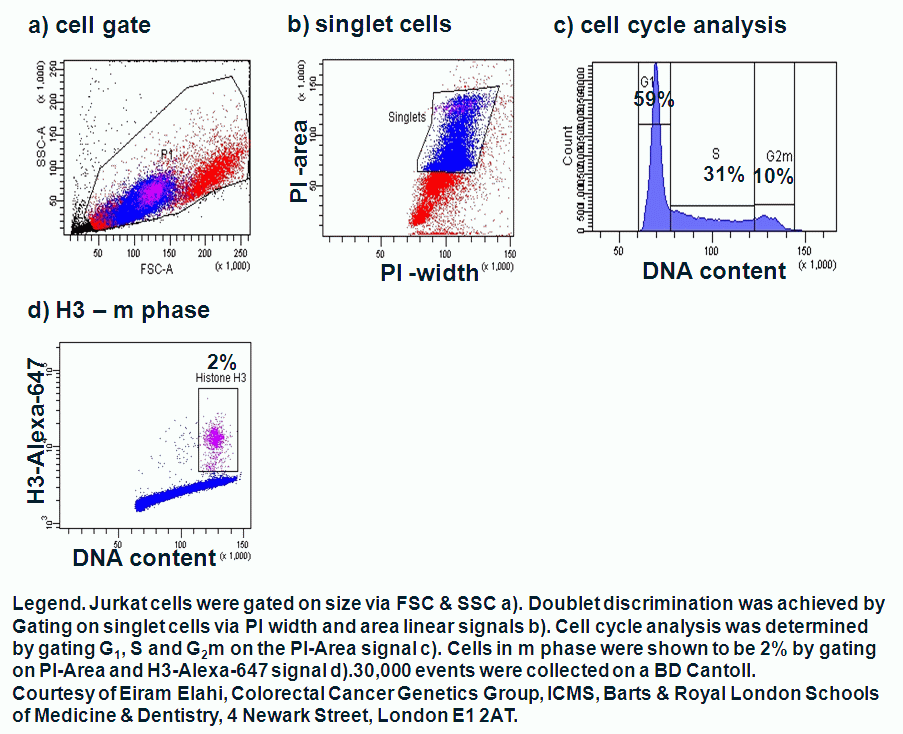
Image - Barts and The London logo and link to home page Image - divider Image - divider
 
  Institute of Cell and Molecular Science
Flow Cytometry Core Facility equipment, images used with permission by BD
  Flow Cytometry Core Facility
  link Home link Instruments available link Flow cytometry link Uses of flow cytometry link Flow sorting link Links link Contacts
 

Determination of m phase


Histone H3


Histones are highly basic proteins that complex with DNA to form chromatin. H3 histone Flow cytometry may be used to analyse cell cycle with propidium iodide (PI) giving peaks of G1 and G2m with S phase located between these peaks. Labelling histone H3 with a fluorescently conjugated monoclonal antibody and then staining PI allows the investigator to determine which G2m cells are in the m phase of the cell cycle by flow cytometry.

 
Top
 
m phase analysis
by Gary Warnes. © Queen Mary, University of London 2007
Institute of Cell and Molecular Science, The Blizard Building, Barts and The London School of Medicine and Dentistry, 4 Newark Street, London E1 2AT, UK Tel: +44 (0)20 7882 2483, Fax: +44 (0)20 7882 2200